Foundational Discovery · 1971
First clinical CT scanner (EMI head scanner)
CT (computed tomography)
Before 1971, diagnosing a brain lesion meant choosing between imprecise plain films and genuinely hazardous procedures: pneumoencephalography required withdrawing cerebrospinal fluid and replacing it with air, while carotid angiography carried its own stroke risk. Neurosurgeons operated, in many cases, on little more than clinical localization. That situation changed abruptly when an EMI engineer named Godfrey Hounsfield installed a prototype scanning machine at Atkinson Morley's Hospital in Wimbledon.
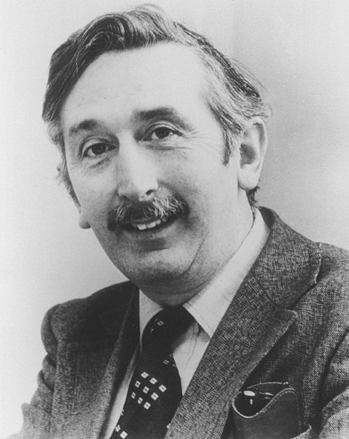
Hounsfield had no medical degree; his background was in radar and computer engineering. Working at EMI Laboratories in Hayes, he reasoned that if a rotating X-ray beam could acquire projections through an object from multiple angles, a computer could reconstruct a cross-sectional image through back-projection. The first clinical scan, performed in 1971 in collaboration with neuroradiologist James Ambrose, imaged a woman with a suspected brain tumor and revealed a frontal lobe cyst with a sharpness no prior technique could provide. The scan took several minutes to acquire and hours more to reconstruct, but the image was unambiguous.
The clinical effect on neurological practice was rapid and thorough. CT could distinguish hemorrhage from ischemic infarction, localize tumors without ventricular contrast studies, and assess hydrocephalus without radiation risk to the spinal cord. Pneumoencephalography, one of the more uncomfortable investigations in neurology, was largely abandoned within a few years of CT's introduction for any indication the scanner could address. Emergency departments reorganized their stroke and trauma pathways around access to the machine.
Hounsfield had not derived the mathematical framework himself; that credit belonged to physicist Allan Cormack at Tufts University, who had published the reconstruction theory years earlier without initially drawing much attention from the medical imaging community. The two men worked independently and never collaborated, yet their contributions were inseparable. In 1979 they shared the Nobel Prize in Physiology or Medicine. Cormack reportedly learned of his prize from a neighbor who heard it on the radio.
Body CT scanners entered clinical use within a few years of the head scanner, extending cross-sectional imaging to the thorax, abdomen, and musculoskeletal system. The Hounsfield unit, the standardized scale for CT attenuation values, is named in his honor and remains the basis for all CT interpretation today. By the time Hounsfield died in 2004, CT scanning had become one of the most performed diagnostic procedures in medicine worldwide.
Key People
- Godfrey Hounsfield — EMI engineer who designed and built the first clinical CT scanner.
- Allan Cormack — Physicist who developed the mathematical reconstruction theory underlying CT imaging.
- James Ambrose — Neuroradiologist at Atkinson Morley's who conducted the first clinical CT scans.
Br J Radiol, 1973
Related landmarks
- 1973 · Lauterbur's NMR imaging (zeugmatography), origin of MRI (Foundational Discovery)
- 1955 · Sanger's Sequencing of Insulin (Foundational Discovery)
- 2006 · Induced Pluripotent Stem Cells (iPSCs) (Foundational Discovery)
- 1932 · Identification of vitamin C as the antiscorbutic factor (Foundational Discovery)