Foundational Discovery · 1895
Roentgen's discovery of X-rays
Before November 1895, the interior of a living patient was accessible only by cutting. Surgeons relied on surface anatomy, percussion, and palpation to locate fractures, foreign bodies, and internal masses. The physical examination was the diagnostic limit. Wilhelm Roentgen was not working on a medical problem when he made his discovery; he was studying the behavior of cathode rays in his laboratory at the University of Wurzburg, using a Crookes tube wrapped in black cardboard to prevent visible light from escaping.
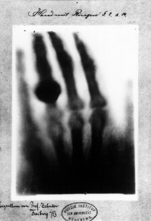
On the evening of November 8, 1895, Roentgen noticed that a screen coated with barium platinocyanide, lying on a bench several feet from the shielded tube, fluoresced when the tube was energized. The cardboard blocked visible light, but something else was passing through it. Over the following weeks he worked in near-total secrecy, characterizing the new radiation: it traveled in straight lines, was not deflected by magnetic fields, passed through soft tissue but was attenuated by bone and metal, and could expose photographic film. He named it X-strahlen, X-rays, because its nature was unknown. On December 22, 1895, he imaged his wife Anna Bertha's hand, capturing bone and her wedding ring on a single photographic plate. She is reported to have reacted with shock upon seeing the skeletal image.
Roentgen submitted his paper, "Uber eine neue Art von Strahlen" (On a New Kind of Rays), to the Wurzburg Physical-Medical Society on December 28, 1895. He sent reprints with sample radiographs to colleagues across Europe in early January 1896. The response was immediate and public: within weeks, newspapers in Vienna, London, and New York had reported the discovery, and the first clinical X-ray images of fractures and foreign bodies appeared in the medical literature within months.
The speed of adoption had no precedent in the history of diagnostic technology. By the end of 1896, X-ray apparatus was in use in hospitals in Europe, North America, and Australia. The military utility was obvious from the start; X-ray units were deployed in the Greco-Turkish War of 1897 and the Spanish-American War of 1898 to locate bullets and shrapnel in wounded soldiers. The hazards of ionizing radiation were not yet understood, and early radiologists and physicists suffered significant radiation injuries before protective practices developed.
Roentgen received the first Nobel Prize in Physics in 1901. He refused to patent the discovery and assigned his prize money to the University of Wurzburg. The technology he described introduced a new category of clinical information: the anatomical image of the living patient. Fluoroscopy, angiography, and computed tomography all descend from the November 1895 observation, as does the entire enterprise of interventional radiology.
Key People
- Wilhelm Roentgen — German physicist who discovered X-rays and described their imaging properties in 1895
- Anna Bertha Roentgen — Roentgen's wife, subject of the first radiographic image of a human hand
Sitzungsber Phys-Med Ges Wurzburg, 1895
Related landmarks
- 1901 · Discovery of the ABO Blood Groups (Foundational Discovery)
- 1912 · Funk's vitamine (vital amine) hypothesis (Foundational Discovery)
- 1861 · Pasteur's refutation of spontaneous generation (Foundational Discovery)
- 1932 · Identification of vitamin C as the antiscorbutic factor (Foundational Discovery)